2024-6-27 05:0:12 Author: hackernoon.com(查看原文) 阅读量:5 收藏
Authors:
(1) Kotaro Shimizu, Department of Physics, University of Virginia, Charlottesville, Virginia 22904, USA and Department of Applied Physics, The University of Tokyo, Tokyo 113-8656, Japan;
(2) Vinicius Yu Okubo, Dept. Electronic Systems Engineering, Polytechnic School, University of São Paulo, Brazil;
(3) Rose Knight, Department of Physics, University of Virginia, Charlottesville, Virginia 22904, USA;
(4) Ziyuan Wang, Department of Physics, University of Virginia, Charlottesville, Virginia 22904, USA;
(5) Joseph Burton, Department of Physics, University of Virginia, Charlottesville, Virginia 22904, USA;
(6) Hae Yong Kim, Dept. Electronic Systems Engineering, Polytechnic School, University of São Paulo, Brazil;
(7) Gia-Wei Chern, Department of Physics, University of Virginia, Charlottesville, Virginia 22904, USA;
(8) B. S. Shivaram, Department of Physics, University of Virginia, Charlottesville, Virginia 22904, USA.
We present a comprehensive approach to characterizing labyrinthine structures that often emerge as a final steady state in pattern forming systems. We employ machine learning based pattern recognition techniques to identify the types and locations of topological defects of the local stripe ordering to augment conventional Fourier analysis. A pair distribution function analysis of the topological defects reveals subtle differences between labyrinthine structures which are beyond the conventional characterization methods. We utilize our approach to highlight a clear morphological transition between two zero-field labyrinthine structures in single crystal Bi substituted Yttrium Iron Garnet films. An energy landscape picture is proposed to understand the athermal dynamics that governs the observed morphological transition. Our work demonstrates that machine learning based recognition techniques enable novel studies of rich and complex labyrinthine type structures universal to many pattern formation systems.
Labyrinthine structures are ubiquitous in out-ofequilibrium nonlinear systems ranging from biological and chemical reactions to fluid convection, crystal growth, and magnetic ordering [1–5]. In such pattern forming systems, the complex structures emerge as a result of energy injection and dissipation in a highly nonlinear process. The labyrinthine patterns are generally characterized by stripe domains of different orientations, sizes, and grain-boundary structures. The predominance of periodic stripes indicates breaking of translational symmetry locally. Yet, contrary to long-range ordered states in an equilibrium phase transition, labyrinthine patterns are essentially disordered and cannot be described by a well-defined order parameter. Indeed, labyrinthine structures can be viewed as an intermediate between featureless short-range correlated glassy state and long-range ordered stripe or crystalline phases [6].
Despite their prevalence in pattern forming systems, a complete characterization of labyrinthine structures is still lacking [7]. A defining characteristic of labyrinthine patterns is the ring-like feature in its structure factor obtained from conventional Fourier analysis [8]. The radius and width of the ring correspond to the wavelength of local stripes and characteristic size of stripe domains, respectively [8–11]. While such global Fourier analysis provides a basic characterization of labyrinths, it fails to capture subtle differences of labyrinthine patterns which have important structural or dynamical implications. Other useful measures, such as the disorder functions [12–14], have been introduced to quantify deviations from a perfect stripe order. Another important characterization often employed is the density of topological defects of labyrinthine structures [15–18]. Indeed, the distribution and correlation between topological defects, such as disclinations and dislocations, of the stripe order encode important information about the labyrinths [17, 18]. However, efficient and accurate identifications of such point-like defects in large-scale experimental or simulation data remain a challenging task.
In this paper, we present a comprehensive framework for the characterization of labyrinthine structures by combining Fourier analysis with machine learning (ML) based template recognition methods. A two-step algorithm that consists of rotation-invariant template matching followed by convolutional neural network analysis was developed to precisely identify topological defects (junctions and terminals) and their coordinates in the images. The high-precision real-space configuration data allow us to compute pair distribution functions of the topological defects, which provide valuable information about labyrinthine patterns that are complementary to those extracted from structure factors.
We apply our approach to studying an intriguing nonequilibrium morphological phase transition in the magnetic medium Ytrium Iron Garnet (YIG), a well known technologically important ferromagnetic material. Magnetic interactions of YIG are dominated by a shortrange ferromagnetic exchange and long-range dipolar interactions [19]. YIG films doped with bismuth are known to carry a strong perpendicular magnetic anisotropy and form complex labyrinthine patterns [16–18]. Bismuth doping introduces a number of advantages desirable for magneto-optic isolators and sensors [20, 21], enhances the Faraday rotation, improves the perpendicular anisotropy and also lowers the saturation magnetization [19–22]. For films grown under appropriate conditions, the magnetization can be saturated in fields less than 100 Oe. Thus the labyrinthine stripe domain patterns are easily observed with a small electromagnet in polarized light under a microscope [23].
Specifically, here we report an intriguing pattern

transition in "annealing" experiments performed on YIG films where a perpendicular magnetic field is stepped down to zero starting from a fully saturated state. The final zero-field state is reached with each subsequent step reduced from the previous following an exponential decay pattern. These domain annealing experiments are carried out with a specific protocol. Starting from the sample in the fully magnetized state, we instantly drop the field to zero where it is held for 10 seconds during which an image is acquired. Further sequential de-magnetization is carried out but with the height of each step reduced exponentially and with the reversed field directions as indicated in Fig. 1(a). All measurements reported here were performed at room temperature. The images obtained covered an area of 2 mm × 1.8 mm. Two samples grown under similar conditions were studied.
Representative images obtained during the beginning of the protocol and at the end of the protocol, are shown in Fig. 1(b) and Fig. 1(c), respectively. Both of them exhibit the labyrinthine stripe patterns and consist of a plethora of defects depicted in Fig. 1(c). These defects where the dark domain ends and three domains meet are termed terminals (+1/2 disclinations) and junctions (−1/2 disclinations), respectively, and are topological defects associated with rotational symmetry breaking [16]. When introducing one of these defects into perfectly ordered stripes, it inevitably gives rise to the appearance of the other. In other words, the disorder manifests as a disclination pair.
The two (both in zero field) images show labyrinthine structures of similar domains, but appear visually different at the same time, making it challenging to quantify their morphology. For further analysis and discussion, we distinguish the domain patterns in Figs. 1(b) and 1(c) as the quenched and annealed states, respectively. In the quenched state, the border of bright and dark domains exhibit a sinuous nature and do not appear as parallel. In contrast, the annealed state consists of significant regions of very parallel domains. This state exhibits roughly equal widths of dark and bright domains, and the areas occupied by them are also roughly equal for any given sample region. This net approximate equality can be achieved by a variety of different patterns which more or less appear the same.


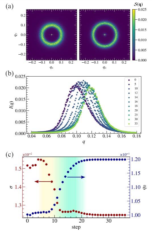
To characterize these morphologically distinct states, we first perform the conventional Fourier analysis by calculating the structure factor

The step dependence of σ and q0 is shown in Fig. 2(c), exhibiting the decrease of σ and the increase of q0 with steps. Notably, σ and q0 exhibit sharp transitions at distinct steps, specifically, in the range of 6 to 12 steps represented by the yellow area and 10 to 20 steps represented by the green area, respectively. It is also clearly seen that when the step height falls below 12 gauss (between steps 10 and 11), the domains settle down to the annealed state. Considering that 1/σ and 2π/q0 correspond to the correlation length and the magnetic modulation period, respectively, we can infer that through the demagnetization process, the labyrinthine structure, which is initially less compact in the quenched state, aligns itself to increase the correlation length and subsequently shortens the magnetic period, transitioning into a more compact annealed state. Application of further smaller steps reorganizes the domain pattern locally at various points but leaves the overall features intac
The conventional analysis conducted thus far provides a certain degree of a global picture in the process of domain compacting. However, it fails to yield a comprehensive understanding of the detailed response of topological defects. As the evolution of the labyrinth patterns is mediated by these defects introduced in Fig. 1, the precise identification of their number and coordinates is inevitable for further clarification.
Nowadays, we have high-resolution digital cameras and much greater computational power compared to what was used in the pioneering work of decades ago [16–18]. Modern popular algorithms for object detection, such as Faster R-CNN [24] and YOLO [25], are based on convolutional neural networks [26]. However, they are not well-suited for defect detection for two reasons. First, they are designed to detect a few large objects, whereas in our context, the goal is to detect thousands of small, closely clustered objects. Second, they require a large number of manually annotated training images, which is extremely labor-intensive to produce due to the high quantity of defects in each image.
We developed a two-step detection algorithm capable of finding thousands of small objects with little manual annotation, which is shown in Fig. 3. First, we employ rotation-invariant template matching to identify candidate points for junctions and terminals [27, 28]. This step is executed with a threshold set to avoid any false negatives, even at the cost of generating several false positives. This approach significantly reduces the annotation workload, as we now only need to review the candidate points rather than locating and classifying every defect in the

image. We selected some images containing candidate points, manually corrected the false positives, and used them to train a convolutional neural network classifier. Therefore, in the second step, the candidate points from the first stage are processed through the classifier, which differentiates between terminals, junctions, and false detections. Manual verification of numerous images indicates that the algorithm’s detection accuracy is nearly 100%. More details about the proposed algorithm will be provided elsewhere.


YIG is renowned for exhibiting a perfect stripe phase in zero magnetic fields (e.g., Fig. 6 of Ref. [18]). However, a labyrinthine structure is observed within the time scale observed in the present study, and even after undergoing a demagnetization process with alternating magnetic fields, the system falls into a more compact labyrinthine pattern without achieving uniform stripe alignment. This behavior implies the presence of a plethora of local minima in the energy landscape, as depicted in Fig. 5. The demagnetization process, by promoting transitions between these local minima, effectively bridges qualitatively distinct quenched and annealed states. In systems governed by such a complex energy landscape, nonlinear responses are expected, and in recent years, they have much attention as a stage for physical reservoir computing. The labyrinthine pattern in magnets, whose complexity can be modulated by an external magnetic field, is expected to be one of the promising venues for physical reservoir computing.
To summarize, we have identified a morphological transition of magnetic domains in a technologically important system and characterized their evolution in new ways by using modern machine learning and pattern recognition analysis. Although similar morphological transitions have been noted before, the present study involves a very subtle transition observed during a demagnetization process. This points to the richness of the magnetic domains in YIG (and other systems) but also is a testimony to the accuracy with which its features can be accurately and quantitatively defined. This has facilitated a detailed analyses about the positional correlation of the defects thus resolving the complex evolution dominating the transition. This is possible due to the high-resolution images and the fidelity of feature recognition and quantification from machine learning tools. These versatile tools will undoubtedly find applications in studies of diverse labyrinthine structures.
We thank Dr. Vincent Fratello and Dr. Shanthi Subramanian of Lucent-Bell Labs for the supply of the LPE grown epitaxial thin films used in this work. We are also grateful to David Huse of Princeton for many helpful comments. The experimental work was partially funded by the NSF under grant DMR and by the 3Cavalier grant from the University of Virginia. This work was also supported by JSPS KAKENHI Grant Number No. JP21J20812. Gia-Wei Chern was partially supported by the US Department of Energy Basic Energy Sciences under Contract No. DE-SC0020330. K.S. was supported by the Program for Leading Graduate Schools (MERITWINGS).
[1] G. Nicolis and I. Prigogine, Self Organization in Non Equilibrium Systems (Wiley, 1977).
[2] M. C. Cross and P. C. Hohenberg, Pattern formation outside of equilibrium, Rev. Mod. Phys. 65, 851 (1993).
[3] A. J. Koch and H. Meinhardt, Biological pattern formation: from basic mechanisms to complex structures, Rev. Mod. Phys. 66, 1481 (1994).
[4] L. Pismen, Patterns and Interfaces in Dissipative Dynamics (Springer, 2006).
[5] M. Cross and H. Greenside, Pattern Formation and Dynamics in Non-Equilibrium Systems (Cambridge University Press, 2009).
[6] M. Le Berre, E. Ressayre, A. Tallet, Y. Pomeau, and L. Di Menza, Example of a chaotic crystal: The labyrinth, Phys. Rev. E 66, 026203 (2002).
[7] S. Echeverría-Alar and M. G. Clerc, Labyrinthine patterns transitions, Phys. Rev. Res. 2, 042036 (2020).
[8] K. R. Elder, J. Viñals, and M. Grant, Dynamic scaling and Quasi ordered states in the two-dimensional swift-hohenberg equation, Phys. Rev. A 46, 7618 (1992).
[9] Q. Ouyang and H. L. Swinney, Transition from a uniform state to hexagonal and striped turing patterns, Nature 352, 610 (1991).
[10] M. C. Cross and D. I. Meiron, Domain Coarsening in Systems Far from Equilibrium, Phys. Rev. Lett. 75, 2152 (1995).
[11] J. J. Christensen and A. J. Bray, Pattern dynamics of Rayleigh-Bénard convective rolls and weakly segregated diblock copolymers, Phys. Rev. E 58, 5364 (1998).
[12] G. H. Gunaratne, R. E. Jones, Q. Ouyang, and H. L. Swinney, An Invariant Measure of Disorder in Patterns, Phys. Rev. Lett. 75, 3281 (1995).
[13] S. Hu, D. I. Goldman, D. J. Kouri, D. K. Hoffman, H. L. Swinney, and G. H. Gunaratne, Stages of relaxation of patterns and the role of stochasticity in the final stage, Nonlinearity 17, 1535 (2004).
[14] S. Hu, G. Nathan, D. J. Kouri, D. K. Hoffman, and G. H. Gunaratne, Statistical characterizations of spatiotemporal patterns generated in the Swift–Hohenberg model, Chaos 15, 043701 (2005).
[15] Q. Hou, S. Sasa, and N. Goldenfeld, Dynamical scaling behavior of the swift-hohenberg equation following a quench to the modulated state, Phys. A: Stat. Mech. Appl. 239, 219 (1997).
[16] M. Seul and R. Wolfe, Evolution of disorder in twodimensional stripe patterns: “smectic” instabilities and disclination unbinding, Phys. Rev. Lett. 68, 2460 (1992).
[17] M. Seul and R. Wolfe, Evolution of disorder in magnetic stripe domains. ii. hairpins and labyrinth patterns versus branches and comb patterns formed by growing minority component, Phys. Rev. A 46, 7534 (1992).
[18] M. Seul, L. R. Monar, and L. O’Gorman, Pattern analysis of magnetic stripe domains morphology and topological defects in the disordered state, Philosophical Magazine B 66, 471 (1992).
[19] P. Hansen and J.-P. Krumme, Magnetic and magnetooptical properties of garnet films, Thin Solid Films 114, 69 (1984), special Issue on Magnetic Garnet Films.
[20] V. Fratello, S. Licht, and C. Brandle, Innovative improvements in bismuth-doped rare-earth iron garnet Faraday rotators, IEEE Trans. Mag. 32, 4102 (1996).
[21] V. J. Fratello, S. E. G. Slusky, C. D. Brandle, and M. P. Norelli, Growth-induced anisotropy in bismuth: Rareearth iron garnets, J. Appl. Phys. 60, 2488 (1986).
[22] T. Fakhrul, S. Tazlaru, L. Beran, Y. Zhang, M. Veis, and C. A. Ross, Magneto-Optical Bi:YIG Films with High Figure of Merit for Nonreciprocal Photonics, Adv. Opt. Mater. 7, 1900056 (2019).
[23] I. B. Puchalska, G. A. Jones, and H. Jouve, A new aspect on the observation of domain structure in garnet epilayers, J. Phys. D: Appl. Phys. 11, L175 (1978).
[24] S. Ren, K. He, R. Girshick, and J. Sun, Faster r-cnn: Towards real-time object detection with region proposal networks (2016), arXiv:1506.01497 [cs.CV].
[25] J. Redmon, S. Divvala, R. Girshick, and A. Farhadi, You only look once: Unified, real-time object detection (2016), arXiv:1506.02640 [cs.CV].
[26] Y. LeCun and Y. Bengio, Convolutional Networks for Images, Speech, and Time Series, in The Handbook of Brain Theory and Neural Networks (MIT Press, Cambridge, MA, USA, 1998) p. 255–258.
[27] H. Y. Kim and S. A. de Araújo, Grayscale TemplateMatching Invariant to Rotation, Scale, Translation, Brightness and Contrast, in Advances in Image and Video Technology: Second Pacific Rim Symposium, PSIVT 2007 Santiago, Chile, December 17-19, 2007 Proceedings (Springer-Verlag, Berlin, Heidelberg, 2007) p. 100–113.
[28] H. Y. Kim, R. H. Maruta, D. R. Huanca, and W. J. Salcedo, Correlation-based multi-shape granulometry with application in porous silicon nanomaterial characterization, J. Porous Mater. 20, 375 (2013).

如有侵权请联系:admin#unsafe.sh